Introduction
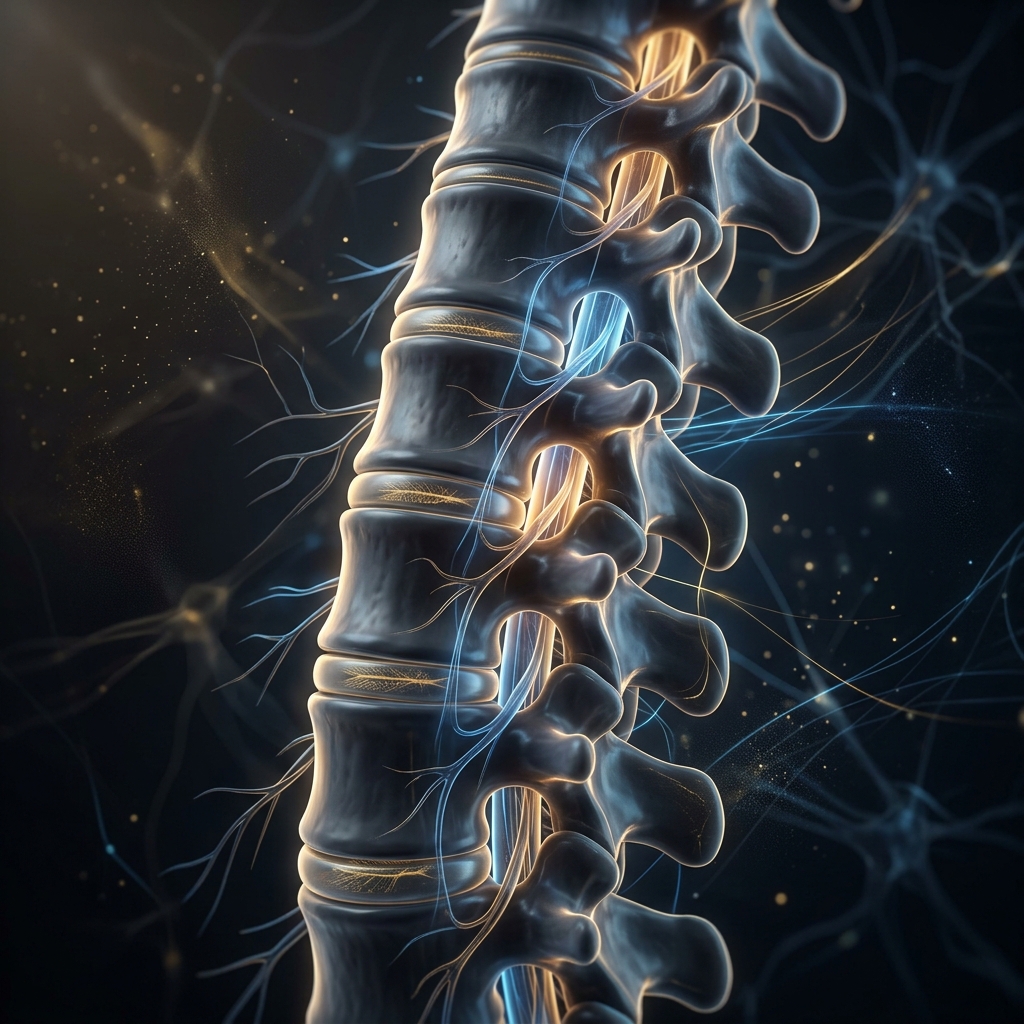
A spinal arachnoid cyst is a benign, fluid-filled sac located within or around the spinal cord, formed by the arachnoid membrane (one of the meninges). These cysts are relatively rare and affect both adults and children. They are most commonly located in the thoracic region, but can also affect the cervical or lumbar spine.
- Intramedullary: Cysts within the spinal cord tissue.
- Extramedullary: Cysts outside the spinal cord but within the arachnoid layer.
Anatomy & Function
The arachnoid is one of the three meninges that cover the spinal cord and brain. The other two meninges are the dura mater (outer) and pia mater (inner). Its primary function is to protect the central nervous system and contain the cerebrospinal fluid (CSF).
Aetiology & Pathophysiology
Spinal arachnoid cysts can be classified into Primary (Congenital) and Secondary (Acquired) causes:
- Developmental Defects: Incomplete separation of the arachnoid membrane during spinal cord development.
- Familial Factors: Some forms have been observed suggesting a hereditary component.
- Acquired Causes: Trauma (spinal cord injury or surgery), Infections (post-infectious arachnoiditis), and certain Spinal Tumors causing mechanical blockage.
Pathologically, fluid accumulates between the arachnoid membrane and the spinal cord. This can disrupt the normal flow of CSF, resulting in increased pressure and leading to severe neurological symptoms, as seen in this clinical case.
Clinical Features & Diagnosis
Symptoms heavily depend on the location and size of the cyst. Common features include chronic back pain, neurological deficits (weakness, numbness in limbs), radiculopathy, and in severe cases, autonomic dysfunction affecting the bowel and bladder.
Diagnosis is primarily established via MRI, which remains the gold standard for revealing the fluid-filled sacs. CT Myelography is also utilized to pinpoint the exact location relative to the spinal cord and nerve roots.
The 14cm Cyst: Case Study
Patient Presentation: A 12-year-old patient suffering from progressive paralysis in both lower limbs with incontinence. Over four years, her condition deteriorated from difficulty in walking to being completely bedridden and unable to sit or turn with her own efforts.
Imaging: MRI spine with contrast revealed severe compression of her dorsal spinal cord on its longitudinal axis. In a growing child, the total spinal cord length is around 40-42cm. The cyst compressing the cord measured an astonishing 14cm in length (from D4 partial to D11 vertebral level).
Surgical Intervention: In a grueling 11-hour marathon surgery, Dr. Khetan meticulously removed the cyst in a single piece from D4 to D11 without rupturing it or damaging the delicate, poorly-vascularized dorsal spinal cord. To prevent long-term bony complications for the young patient, the bone protecting the spinal cord was completely replaced and fixed for fusion (laminoplasty).
Outcome: The patient is gradually improving, now able to sit with her own efforts within a span of 3 months. Histopathology confirmed an Arachnoid cyst. This is claimed as a world record for the longest cyst (14cm) ever removed longitudinally in a growing child.