Overview of Meningiomas
Meningiomas are typically slow-growing tumors that arise from the meninges, the protective membranes surrounding the brain and spinal cord. While usually benign, they can sometimes go undetected for long periods due to their insidious, displacing growth. Over time, this slow expansion gradually displaces healthy surrounding brain tissue, which can lead to severe neurological damage and disability.
Clinical Significance
In rare cases, meningiomas can infiltrate surrounding tissue and metastasize. The localization of intracranial meningiomas is diverse, appearing at the base of the skull, the cranial dome, the falx cerebri, or even intraventricular regions.
Classification & Symptoms
According to the World Health Organization (WHO), meningiomas are classified into three grades of severity based on fine tissue criteria:
- WHO Grade I: Benign and slow-growing (most common).
- WHO Grade II (Atypical): Benign, but grow more rapidly, have an increased tendency to infiltrate surrounding tissue, and often recur after successful therapy.
- WHO Grade III (Anaplastic): Malignant tumors that grow rapidly and always require additional radio-chemotherapy.
Symptoms typically intensify gradually and depend entirely on the tumor's location and growth speed. They often include:
- Epileptic seizures
- Headaches
- Visual, olfactory, and speech disorders
- Paralysis and changes in sensitivity
- Often discovered only as incidental findings
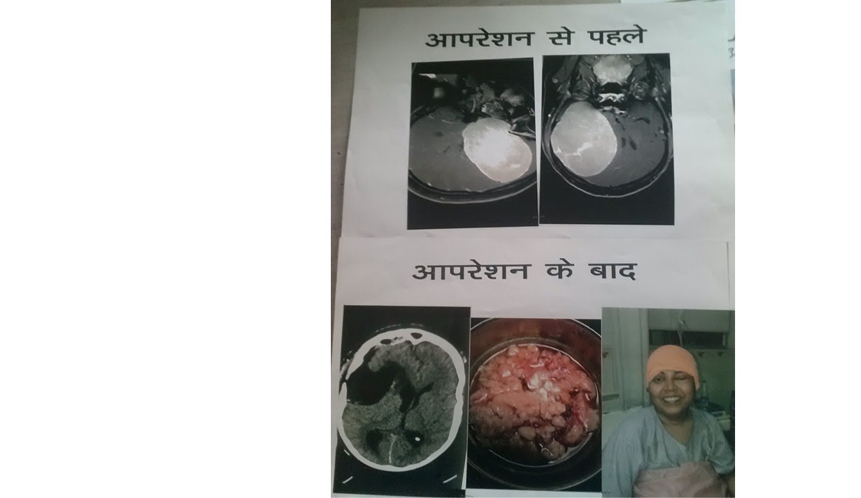
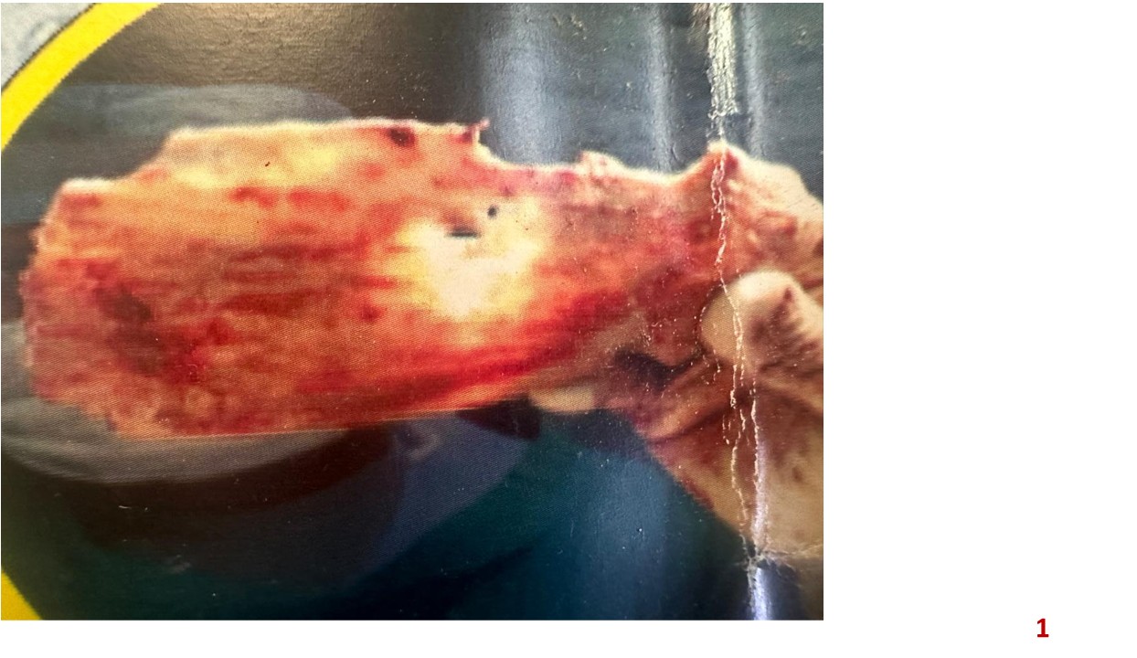
The 240g Tumor: Case Study
Dr. Khetan performed one of the most complicated brain tumor surgeries to date. The patient presented with a massive meningioma situated in an extremely precarious and highly vascularized location.
Surgical Intervention: In a grueling 8-hour marathon surgery, the 240-gram meningioma was meticulously and completely extracted. The surgical challenge was amplified by the tumor's location—lodged tightly between the superior and inferior sagittal sinus, crossing the midline, and actively invading the inferior sagittal sinus.
Outcome: Despite the intense complexity of preserving the critical venous sinuses and surrounding healthy brain tissue, the tumor was removed successfully without neurological deficits, giving the patient a new lease on life.